Meaning Of X-ray Of Chest
Hyperaeration of lungs on a chest xray is caused by abnormal increase in total capacity for air in the lungs. Describing a chest X-ray abnormality can be likened to describing a skin rash in a dermatology patient or a lump in a surgical patient.

Top Chest X Ray Interpretation Books In Amazon Critical Care Practitioner X Ray Medical Textbooks Nuclear Medicine
It is not necessarily an abnormality but often seen in patients with chronic obstructive pulmonary disease copd.

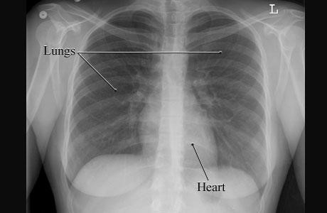
Meaning of x-ray of chest. A chest X-ray doesnt show the inside structures of the heart though. Right middle lobe pneumonias typically obscure the. An X-ray is an imaging test that uses small amounts of radiation to produce pictures of the organs tissues and bones of the body.
An X-ray film is positioned against the body opposite the camera which sends out a very small dose of a radiation beam. It first appears too complicated to read the chest xrays because we barely know what lies where and what to make out of it. An abnormal area of infiltrate on a chest X-ray can represent many abnormalities such as infection water or edema tumor abnormal inflammation not related to infection scarring collapsed lung tissue and other things.
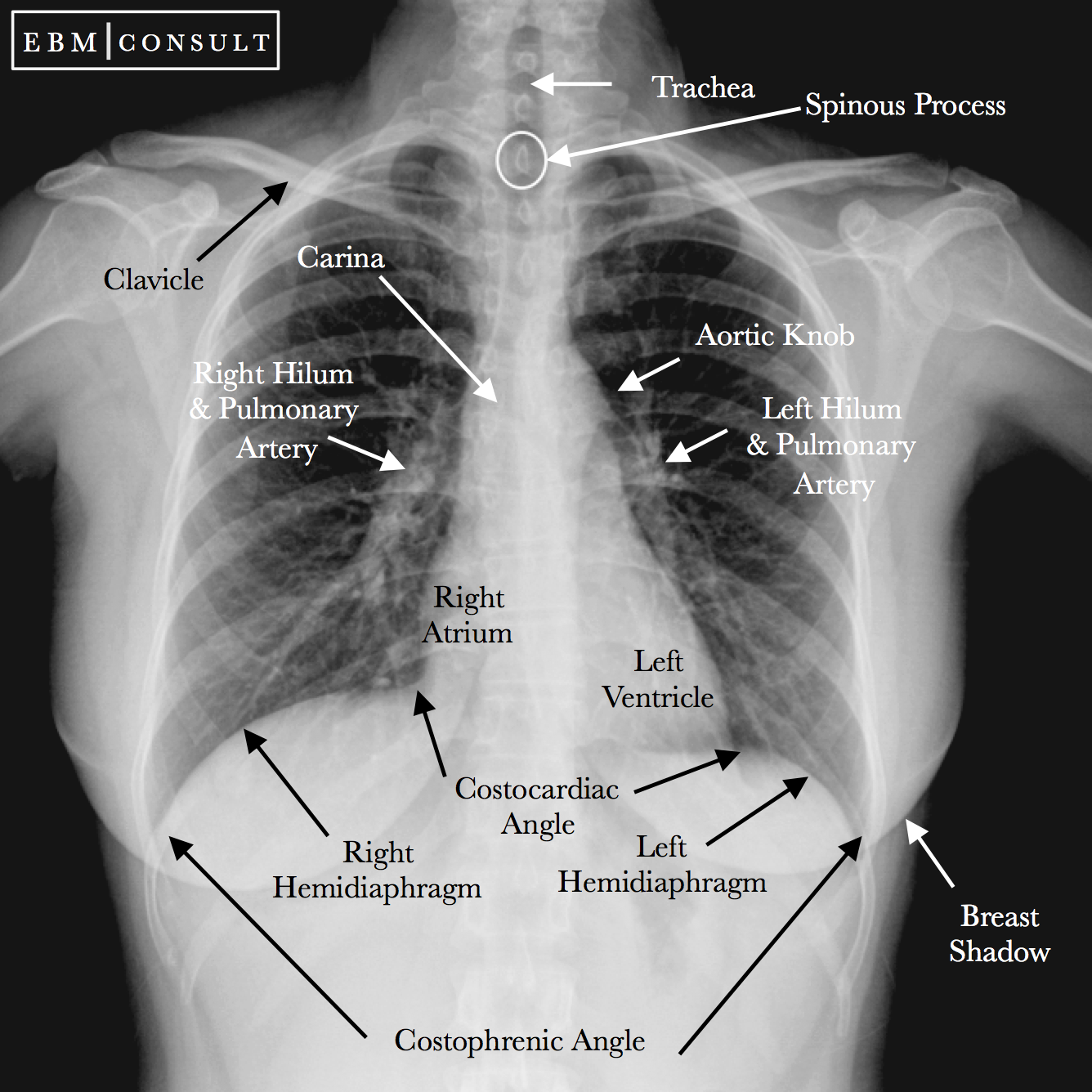
A chest X-ray is a picture of the heart lungs and bones of the chest. Small opacity left base believed to be a confluence of shadows. A chest X-ray shows the location size and shape of the heart lungs and the blood vessels.
This is one of the most common findings on a chest x-ray. Atelectasis is almost always associated with a. Attention should be given to factors such as location size shape and density of an abnormality.
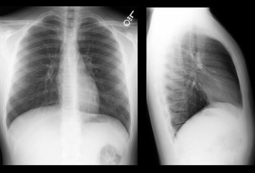
It is most often caused by an endobronchial lesion such as mucus plug or tumor. In medical radiography an X-ray generator produces a beam of energy x-rays that travels towards the body of the patient. Uses of X-Ray Chest PA and LAT View Test The X-Ray examination is done to evaluate any abnormalities in the heart chest wall and lungs.
X-Ray is a type of radiography and most widely used investigation. What does confluence of shadows mean in radiology. Atelectasis is collapse or incomplete expansion of the lung or part of the lung.
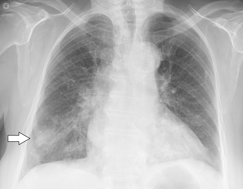
Chest pain shortness of breath and cough are among the myriad reasons chest X. Infiltrate on a chest X-ray report is a common finding that radiologists use to describe a white abnormal area of unclear cause. The absence of lung markings on the right side are used for this diagnosis source Pneumonia.
Chest X-rays or radiographs are one of the most commonly performed imaging studies in all of radiology. The process of description often helps with diagnosis -. Chest x ray.
X ray is a type of radiography and most widely used investigation. Especially good to confirm air-fluid levels in the lung. Why is it done.
This increased air expansion makes the lungs darker on an xray more lucent than normal. A chest X-ray is a radiology test that involves exposing the chest briefly to radiation to produce an image of the chest and the internal organs of the chest. Radiographs commonly known as X Rays are images obtained for diagnostic purposes.
Part of these X rays will be absorbed by body structures while some of them will make it through the body and will be captured on a film placed behind the patient. An X-ray done to detect lung disease or to determine the size and position of the heart ribs or other internal structures of the chest. Basics of Reading Chest X-ray Simply Explained By Medicos Times.
It first appears too complicated to read the chest X-Rays because we barely know what lies where and what to make out of it. A very common lung finding X-rays can be used to definitively diagnose pneumonias. When focused on the chest it can help spot abnormalities or.
But the basics of Chest Xray here will guide you through various aspects including Counting ribs PA vs AP view Inspiratory vs Expiratory Xray Erect vs Supine Lucency and Opacity and some common. The X-ray image is generally the first option to diagnose symptoms like. It is good because it means that there is nothing wrong with you lungs heart size and overall structure of the organs inside your chest cavity - at least from what it can be seen from a chest xray.
Right Anterior Oblique Right Anterolateral Chest Next to Cassette Decubitus Views decubitus actually means lying down made with the patient lying on his side and the x-ray beam horizontal parallel to the floor. Right pneumothorax seen on chest X-ray.
Radiology Chest Xray Normal

How To Read Chest X Rays International Emergency Medicine Education Project

Button Artifact Chest X Ray Radiology Case Radiopaedia Org
![]()
Normal Chest X Ray Anatomy Tutorial Kenhub

Dark Lung Spots On X Ray Lunges Dark Spots Black Lungs
Abnormal Chest X Ray What Is It Symptoms Causes Prevention And Treatment Top Doctors

Chest X Ray Expiration And Inspiration Views Radiology Case Radiopaedia Org

Pulmonary Plethora Causes Obv Left Heart Abnormalities Double Outlet Right Ven Human Anatomy And Physiology Nuclear Medicine Anatomy And Physiology
Cardiomediastinal Outlines On Chest X Ray Radiology Case Radiopaedia Org

Consolidation Density In Left Lower Lung Field Loss Of Left Heart Silhouette Pathology Lunges X Ray

Shoulder E Ray Normal Shoulder X Rays Radiology Case Radiopaedia Org X Ray Anatomy Art Radiology
Chest X Ray St Vincent S Lung Health

Basics Of Reading Chest Xray Complete Guide To Cxr Beginners Medicforyou In 2021 Medical Studies Radiography X Ray

Chest Radiograph Radiology Reference Article Radiopaedia Org

Pin On Devgru Nvd Cnvd Equipments

Pa Vs Ap View Human Anatomy And Physiology Apex Of Lung Radiology
What To Look For On A Chest X Ray Slideshow
Anatomy Of A Chest X Ray How To Read A Chest X Ray Part 1 Youtube

Chest X Ray
Post a Comment for "Meaning Of X-ray Of Chest"